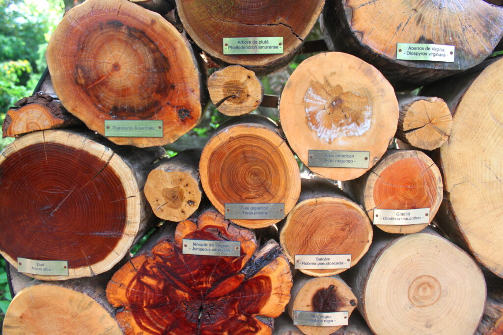
Mulberry – Morus alba Eastern Red Cedar – Juniperus virgiana Western Red Cedar – Thuja plicata Black Locust – Robinia pseudoacacia Phellodendron amurense – Pterocarya fraxinifolia Common Persimmon – Diospyros virgiana Honey Locust – Gleditsia triacanthos Black walnut – Juglans nigra
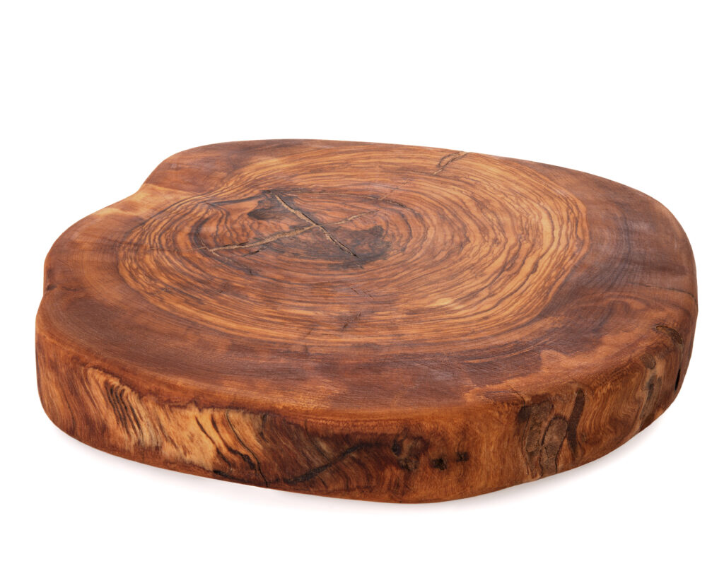
AmbrosiaBirchBirch BirchAshBeechOlivewoodLarchDouglas Fir (BC Fir)Eastern White Pine – Pinus strobusRed Pine – Pinus resinosa Red Pine – Pinus resinosa Douglas Fir – (BC Fir) – Pseudotsuga menziesiiBlack Locust – Robinia pseudoacacia Elm Maple – Acer Saccharum Larch – Tamarack Larch – TamarackGenuine Teak – Tectona grandis Aspen
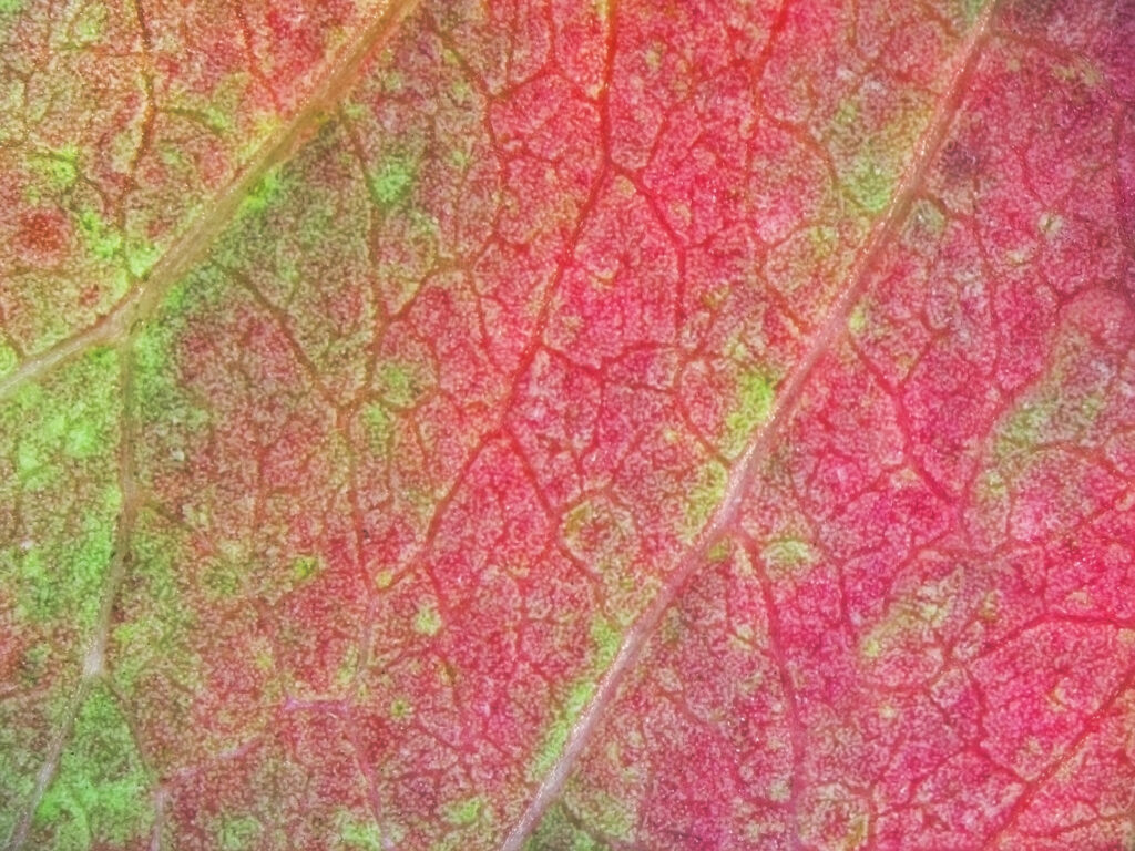
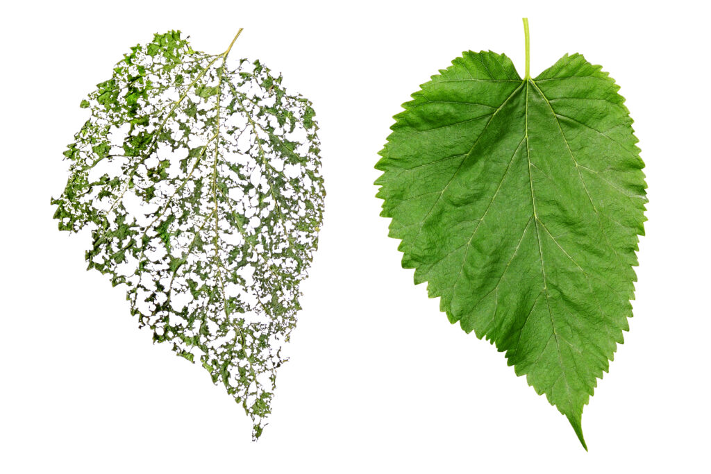
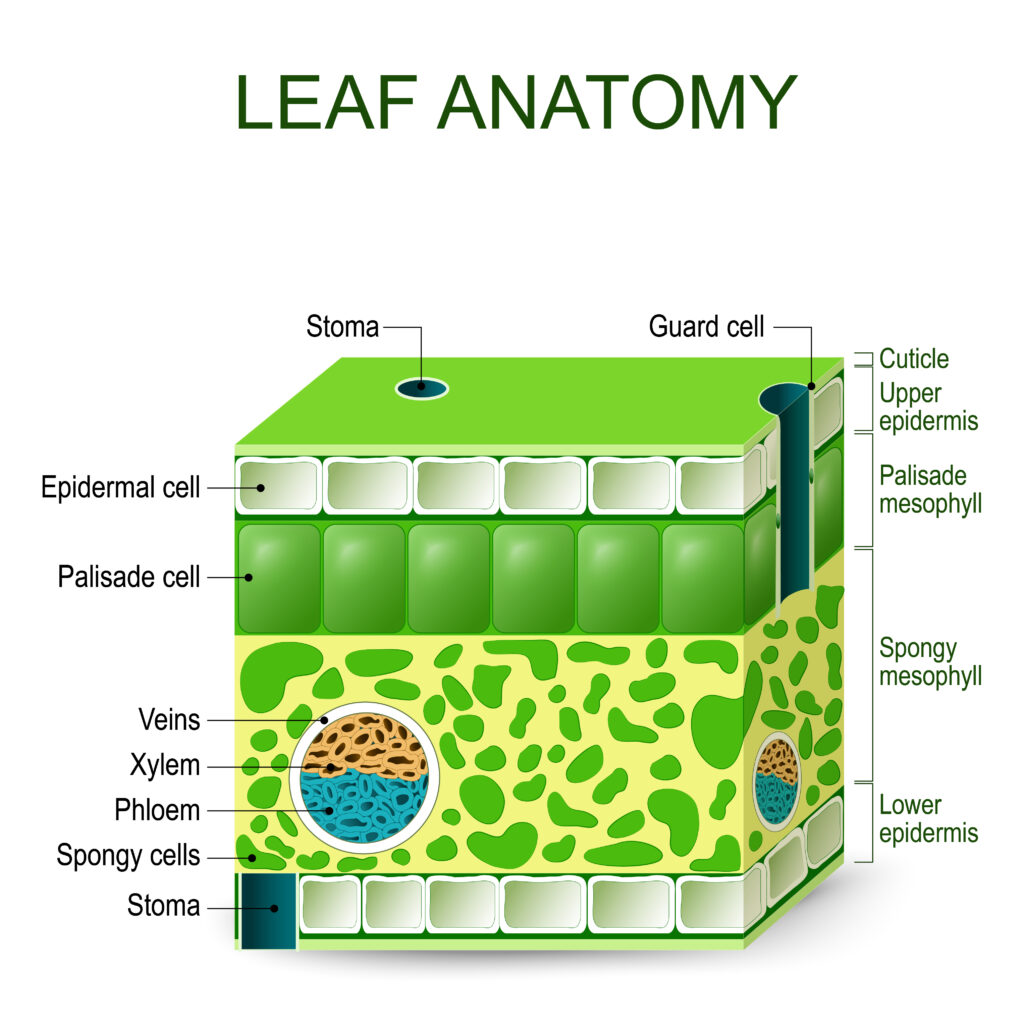
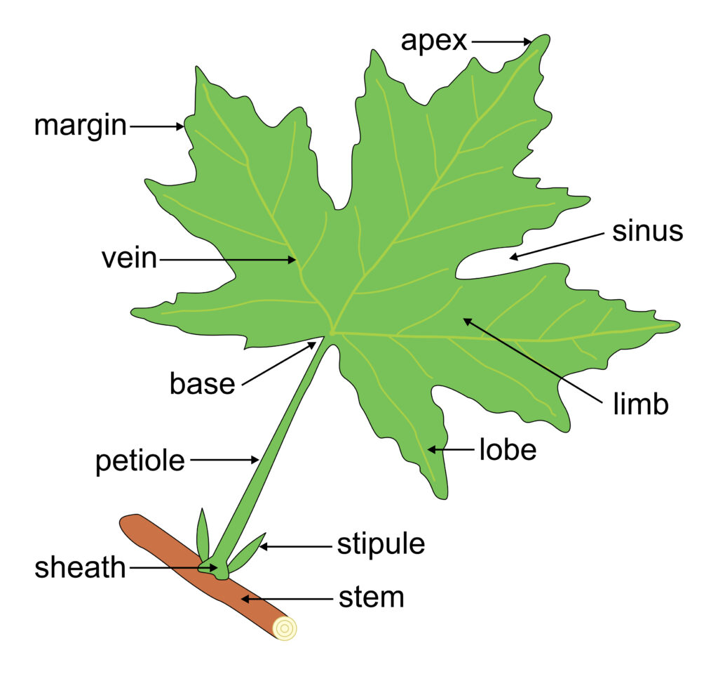
Leaves
Under the microscope
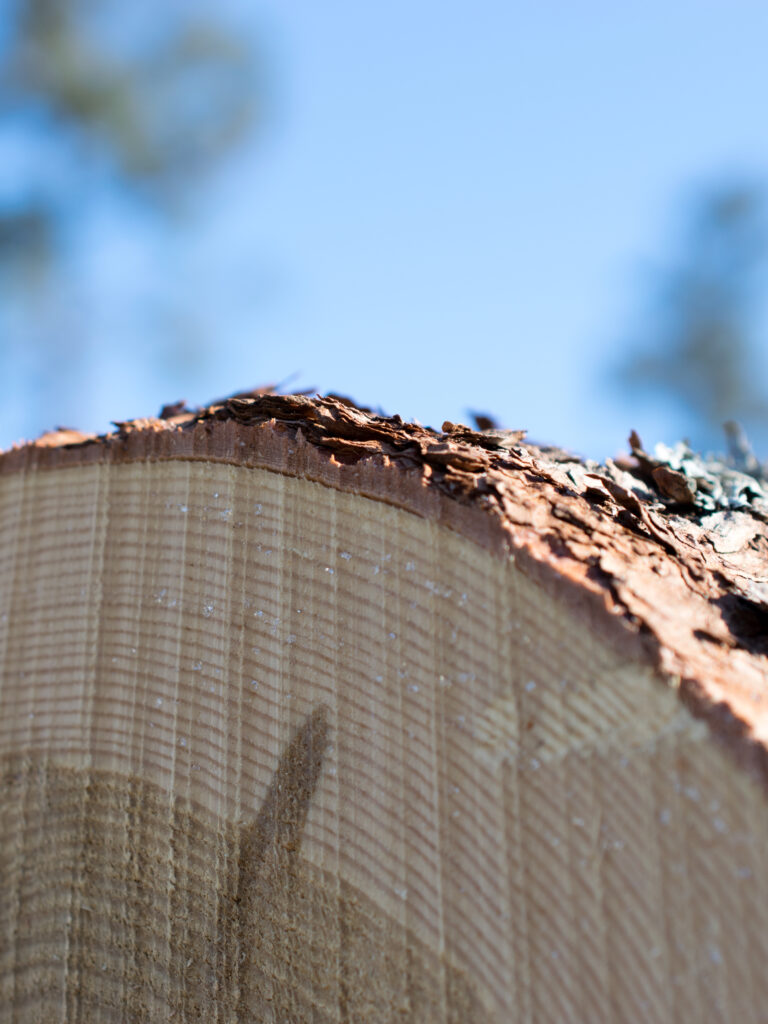
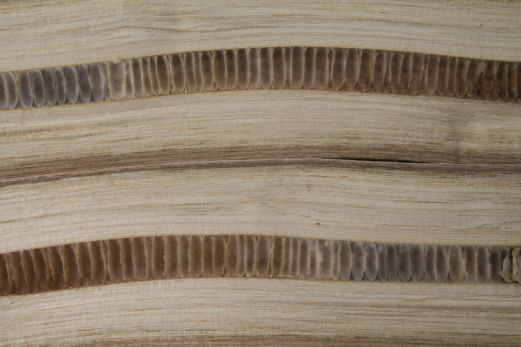
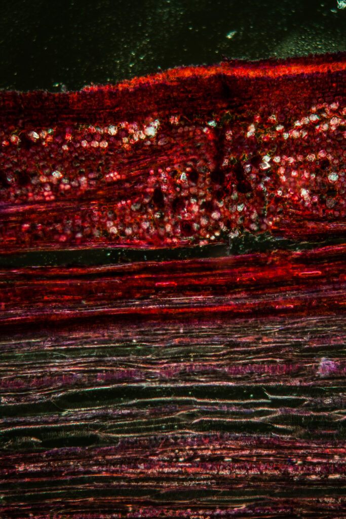
Arteries in the split branch
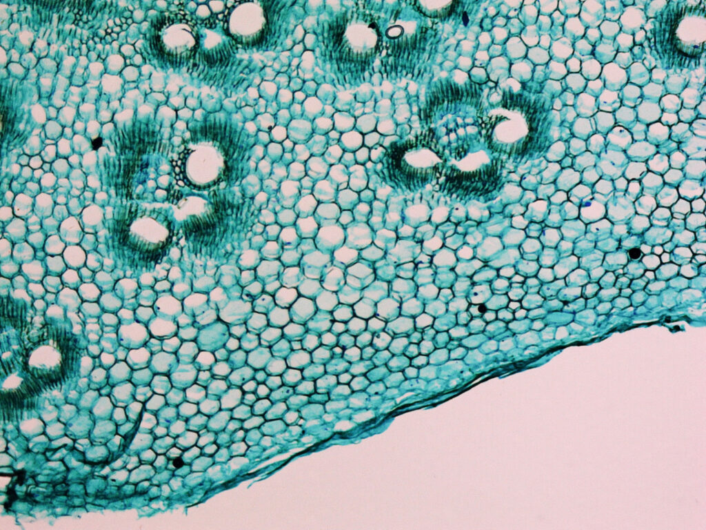
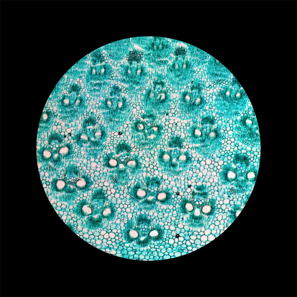
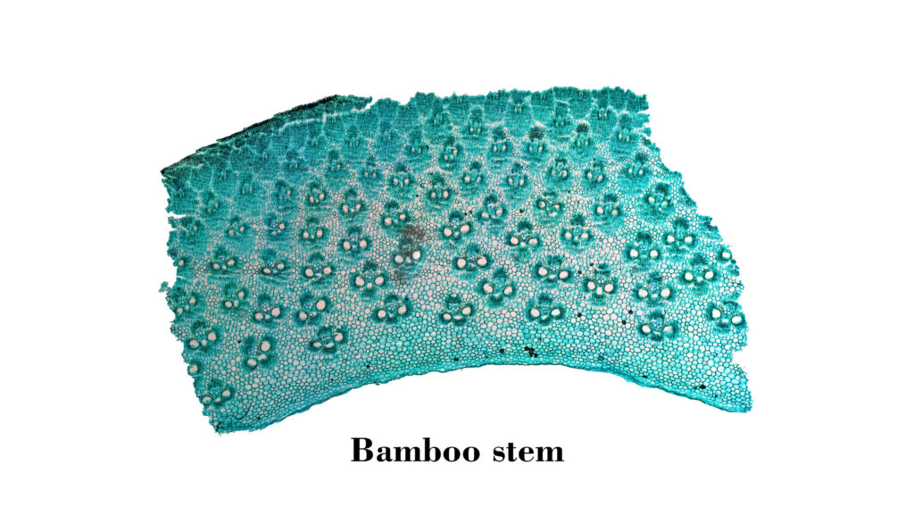
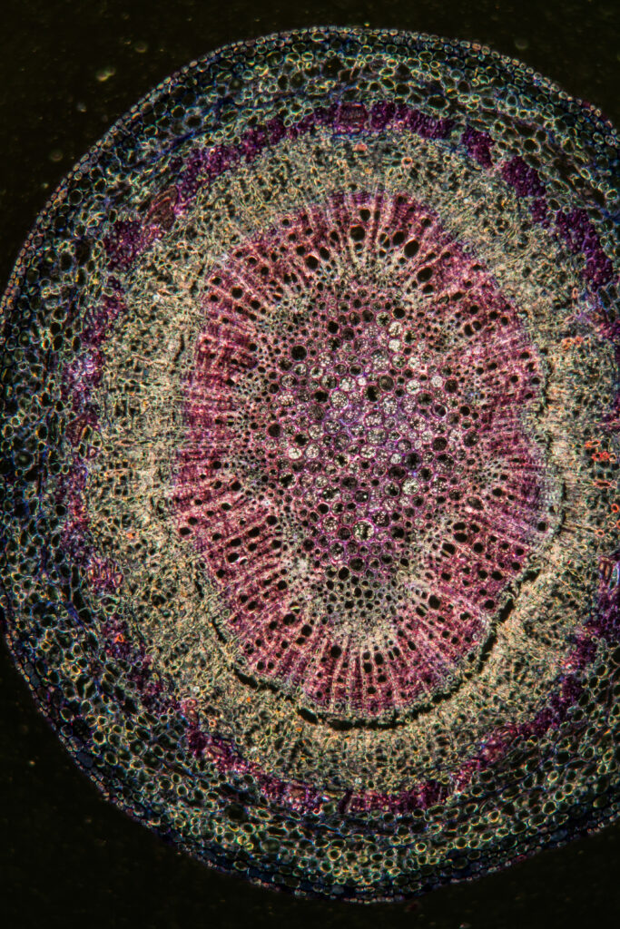
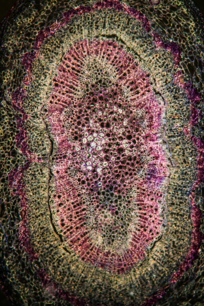
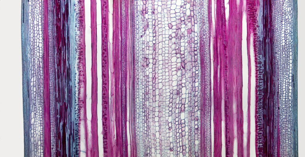
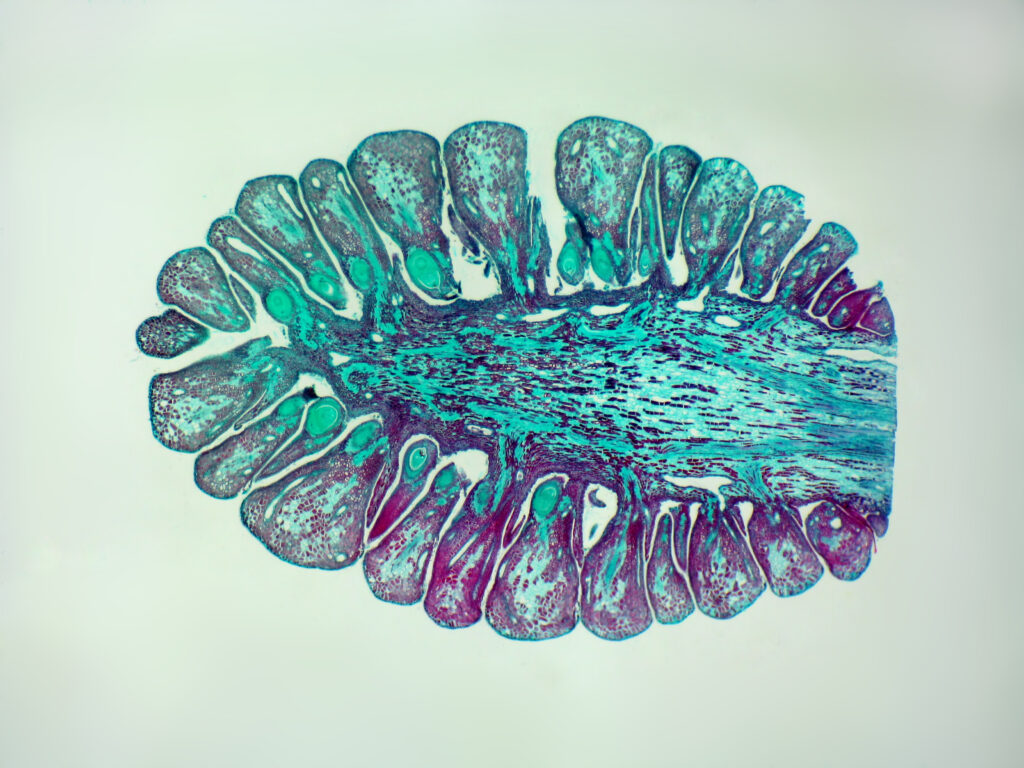
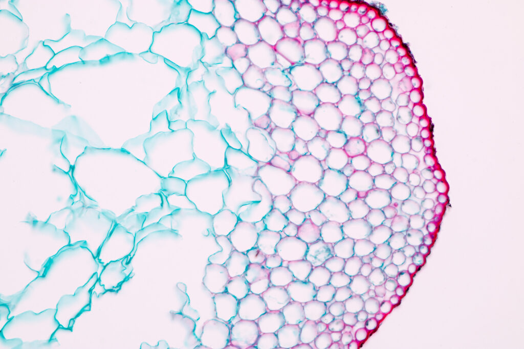
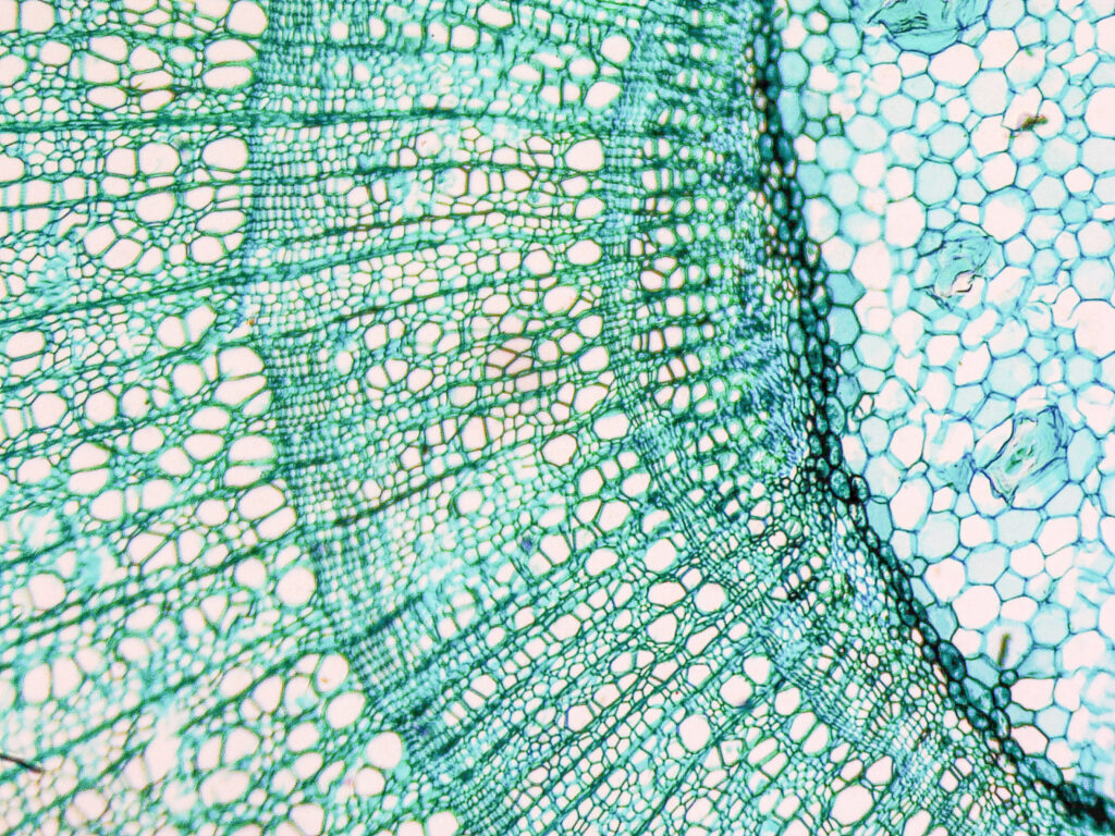
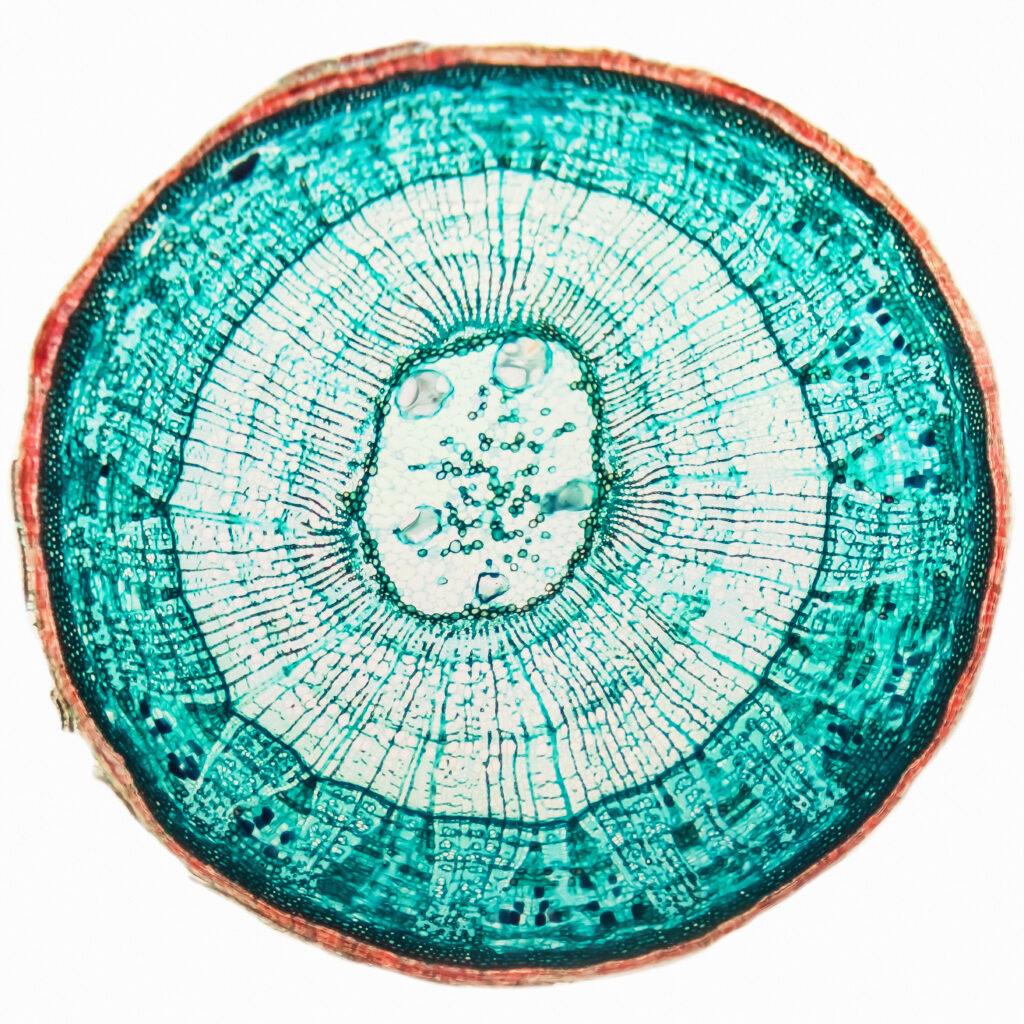
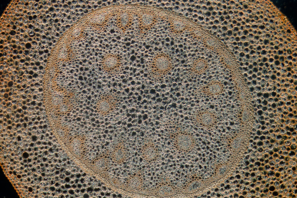
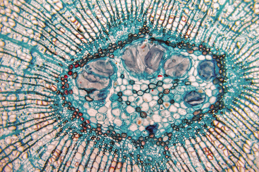
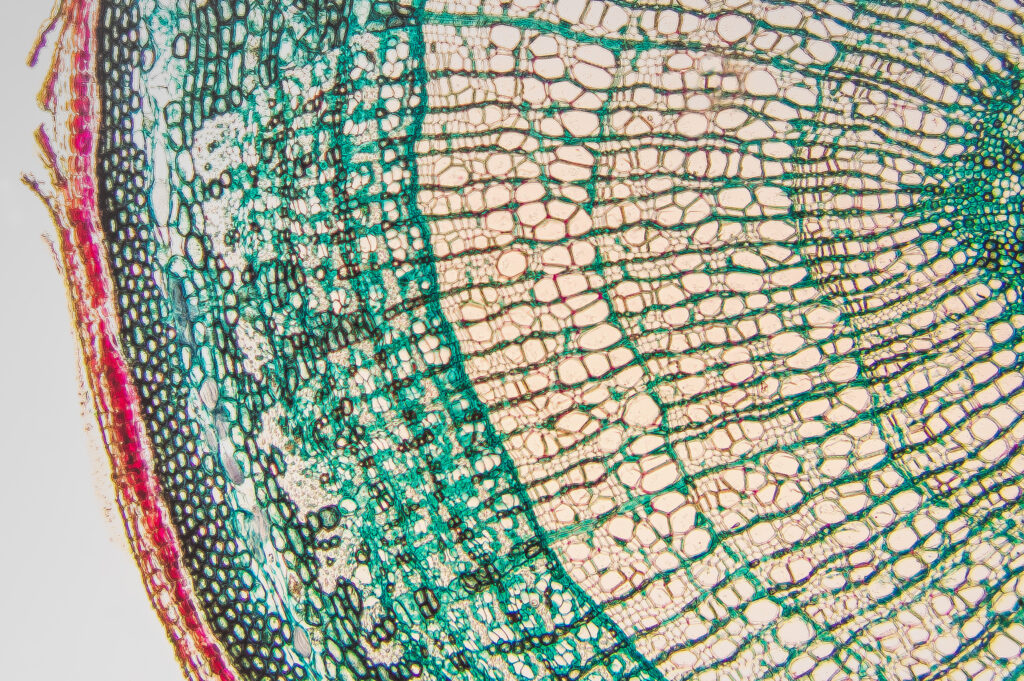
Bamboo
Light photomicrograph of Bamboo Stem cross section seen through microscopeLight photomicrograph of Bamboo Stem cross section seen through microscopeLight photomicrograph of Bamboo Stem cross section seen through microscope
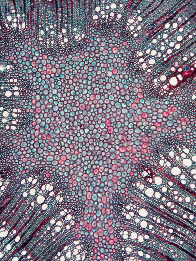
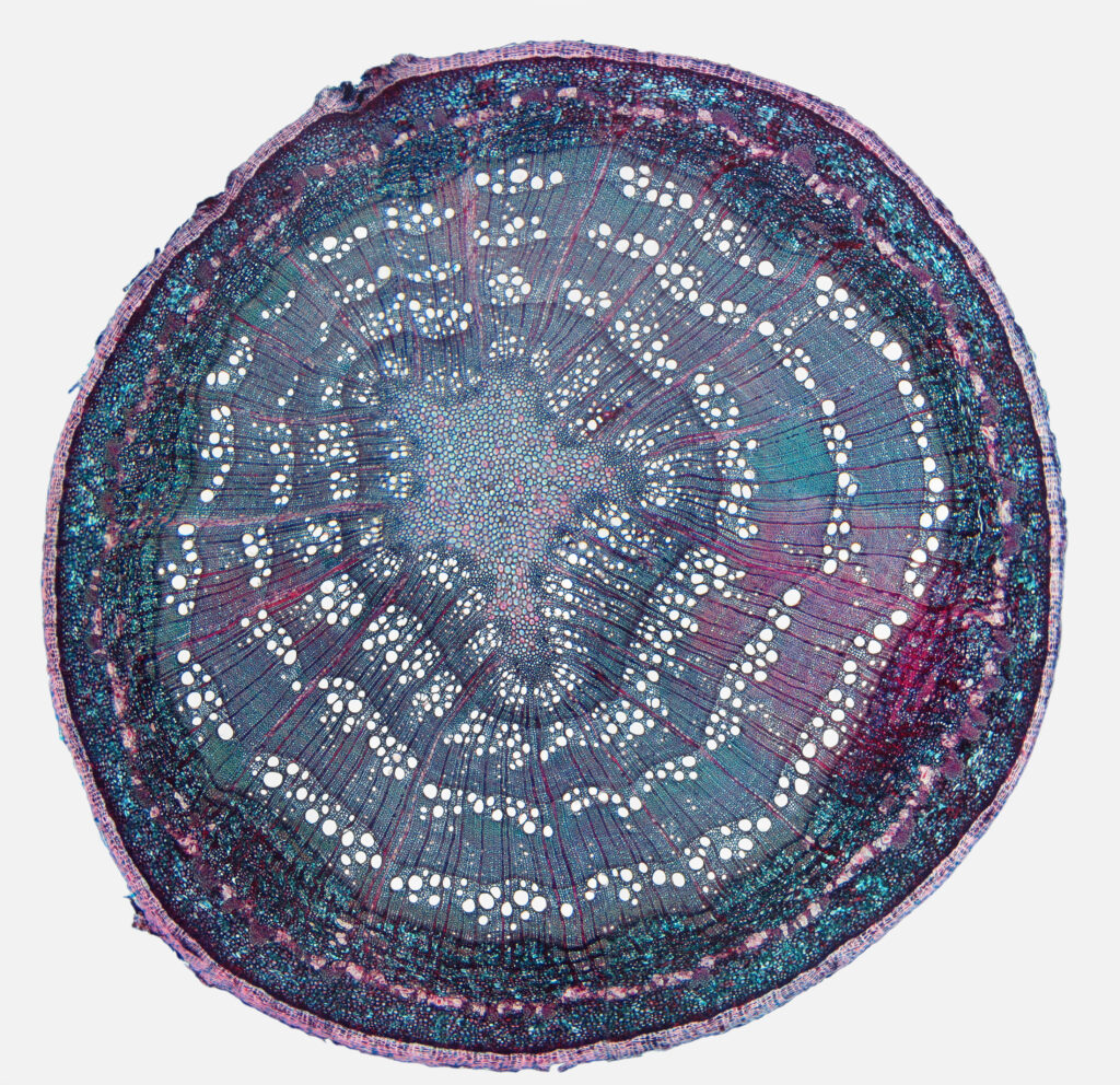
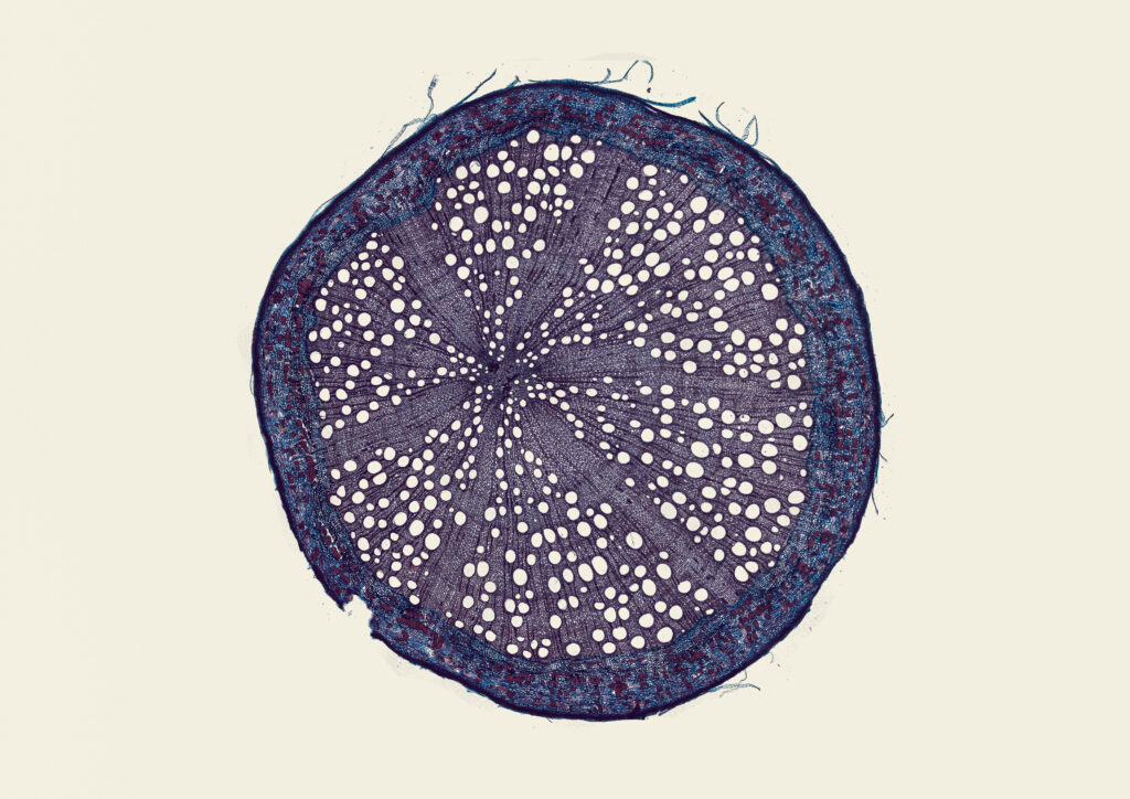
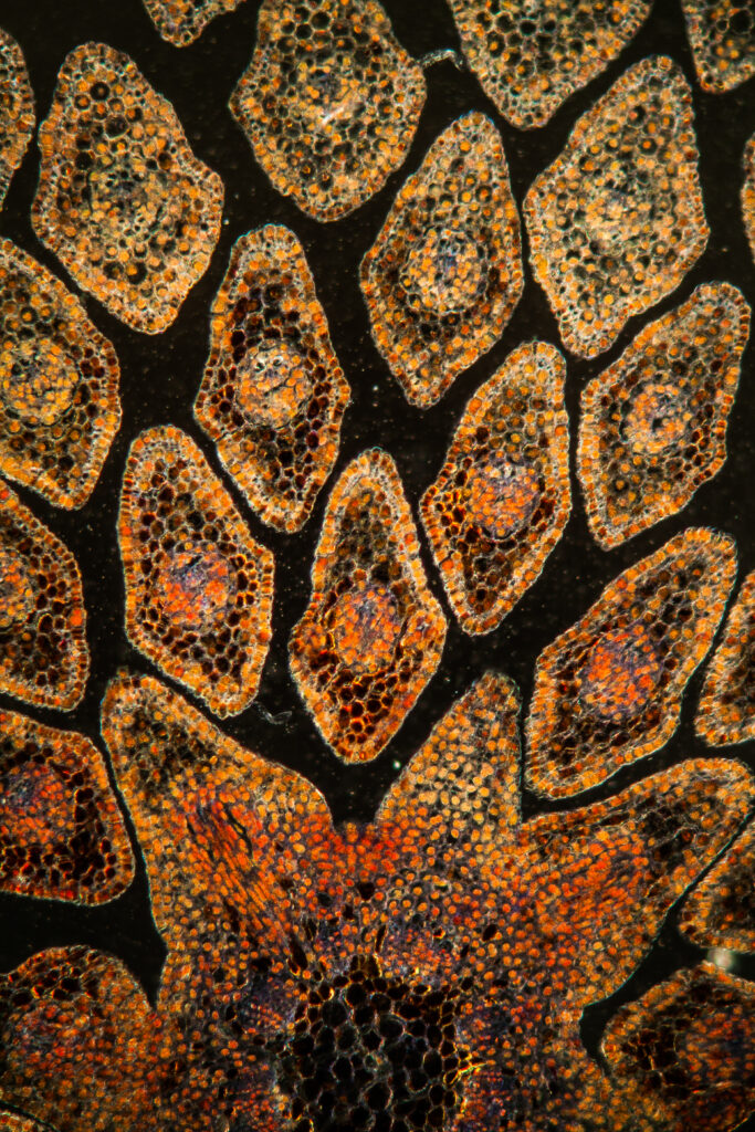
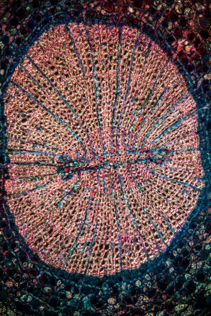
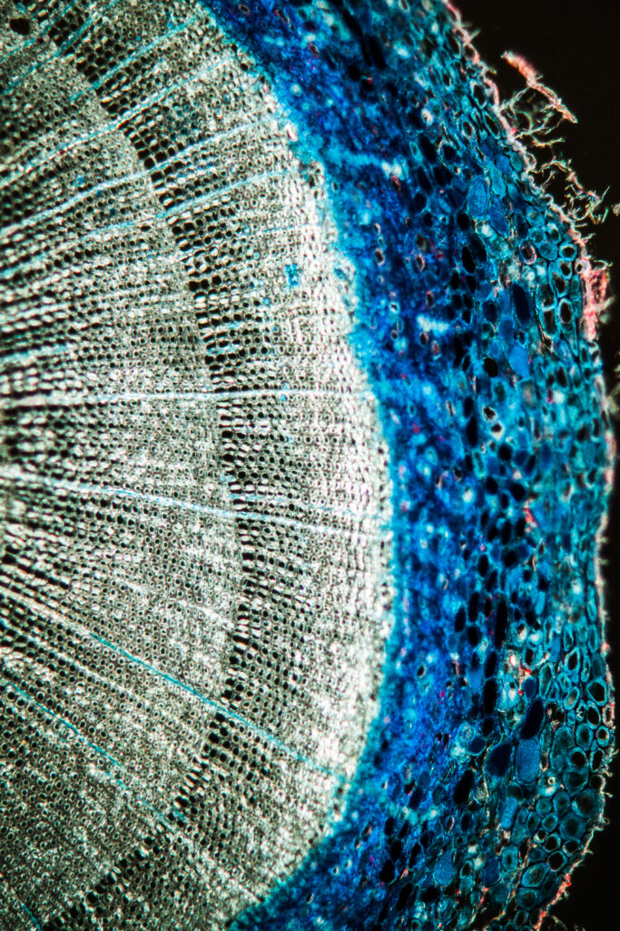
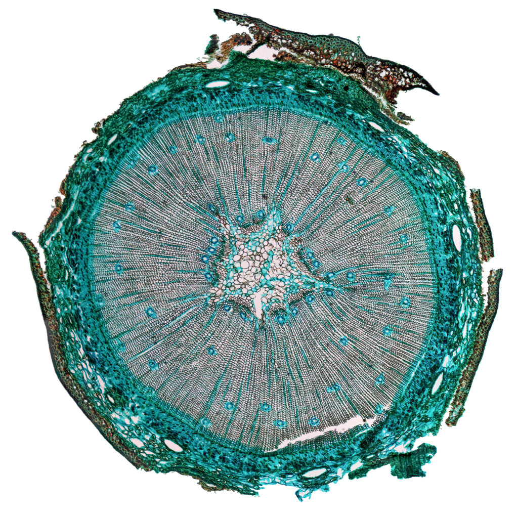
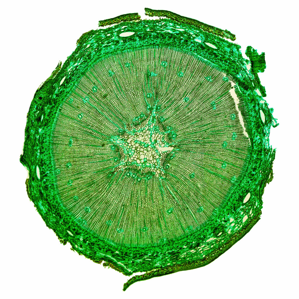
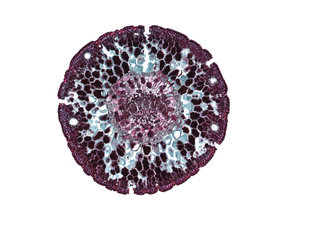
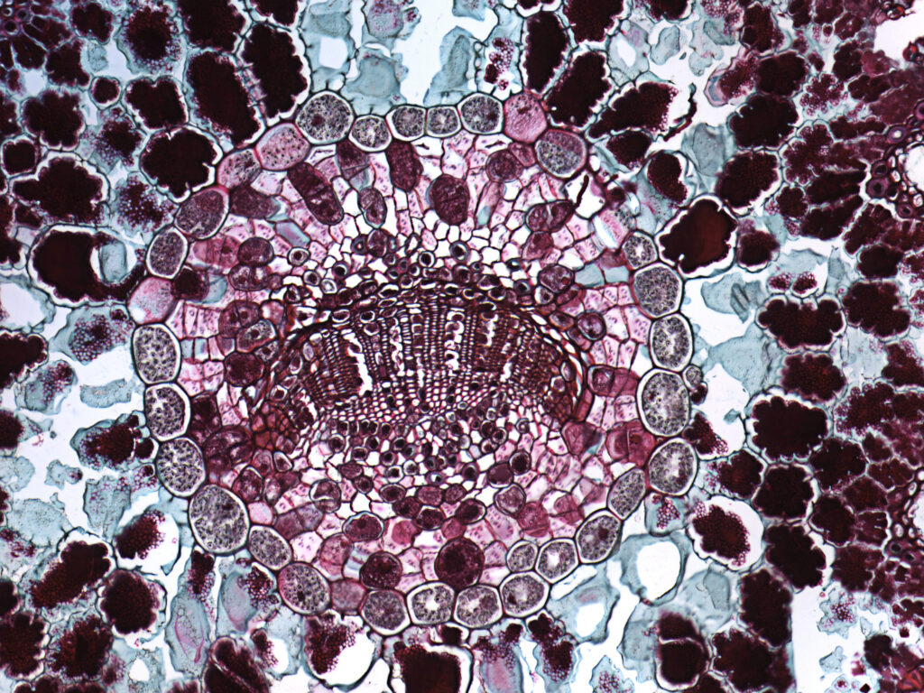
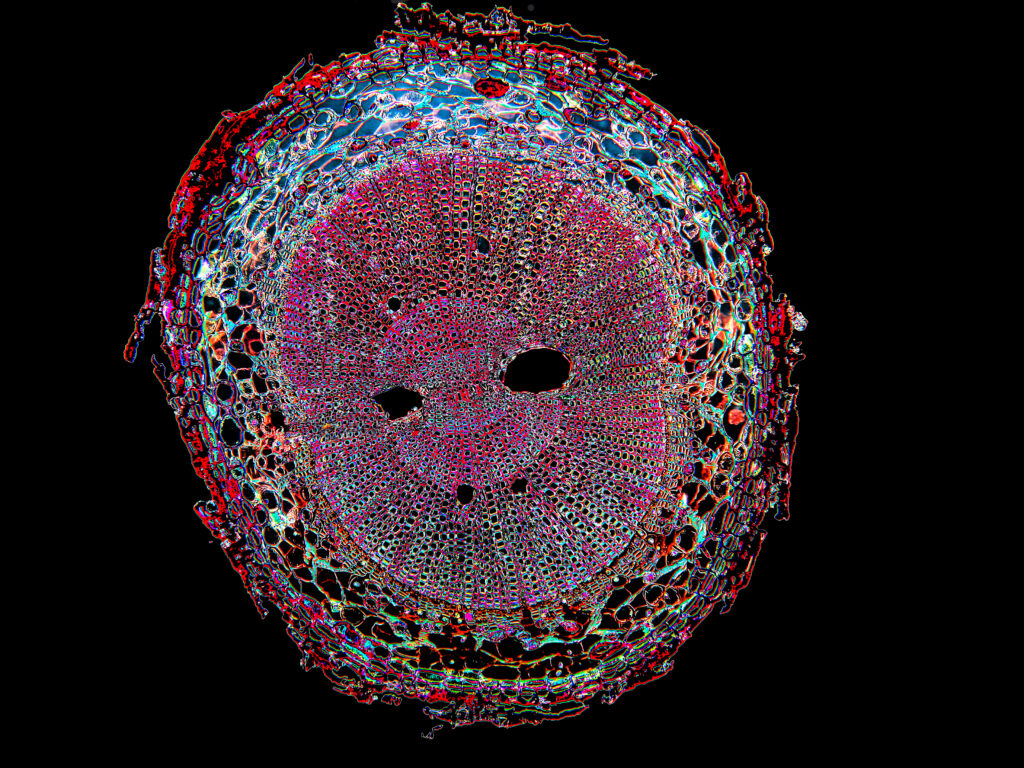
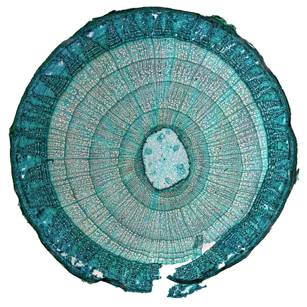
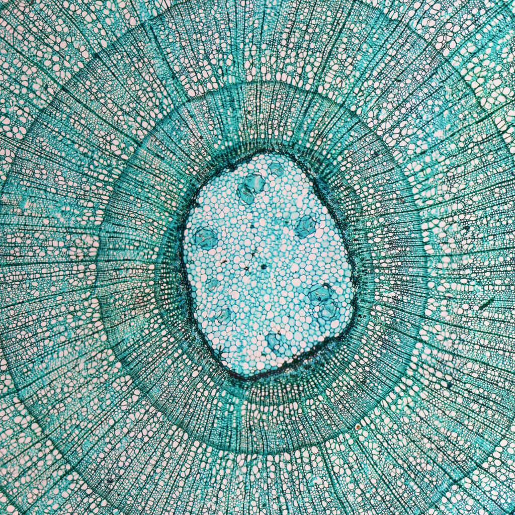
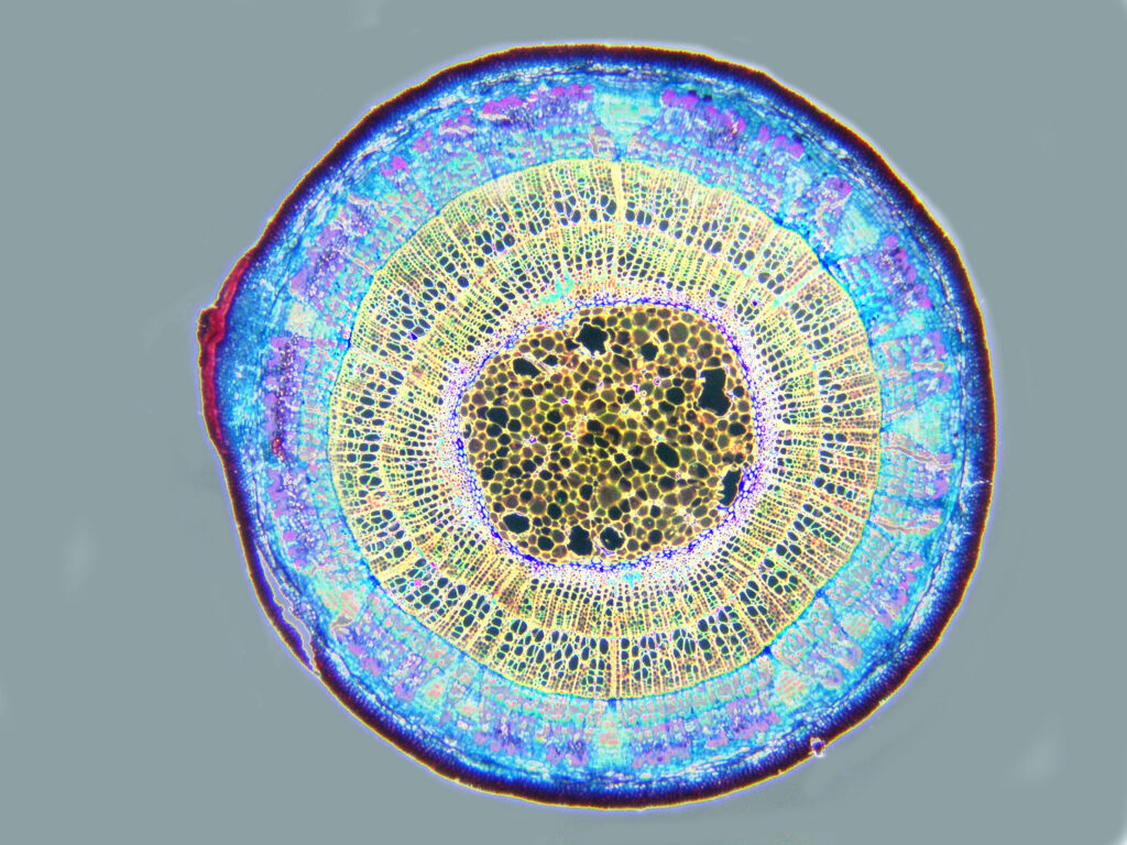
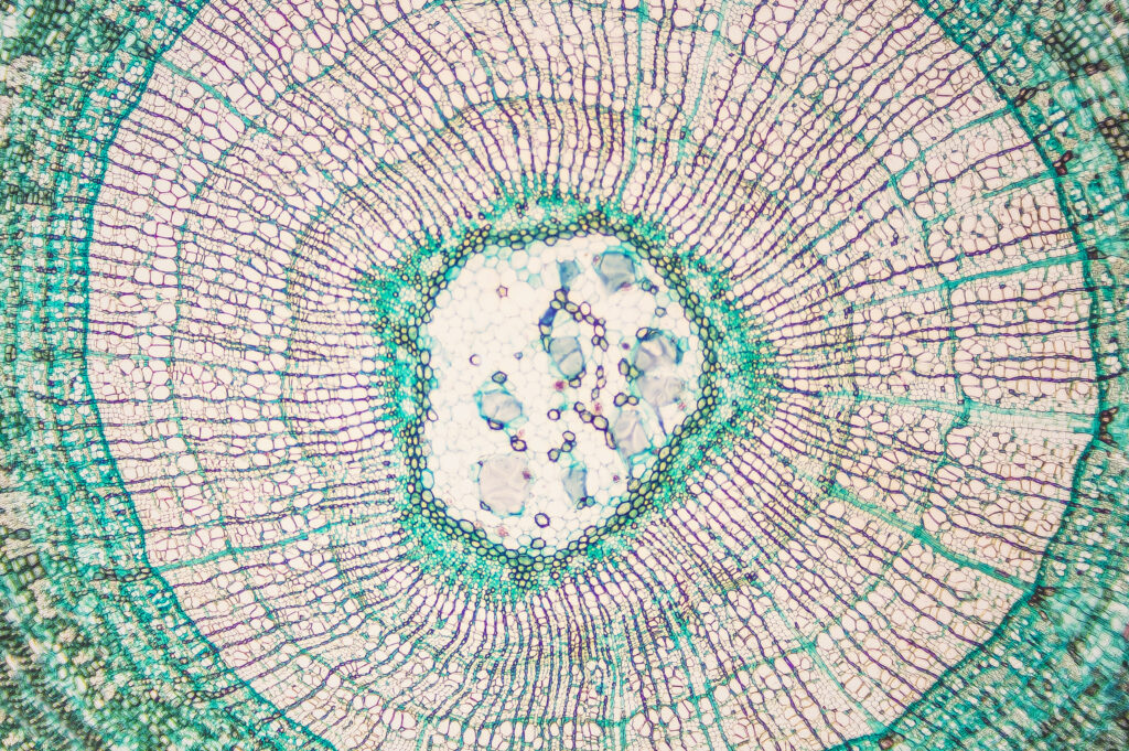
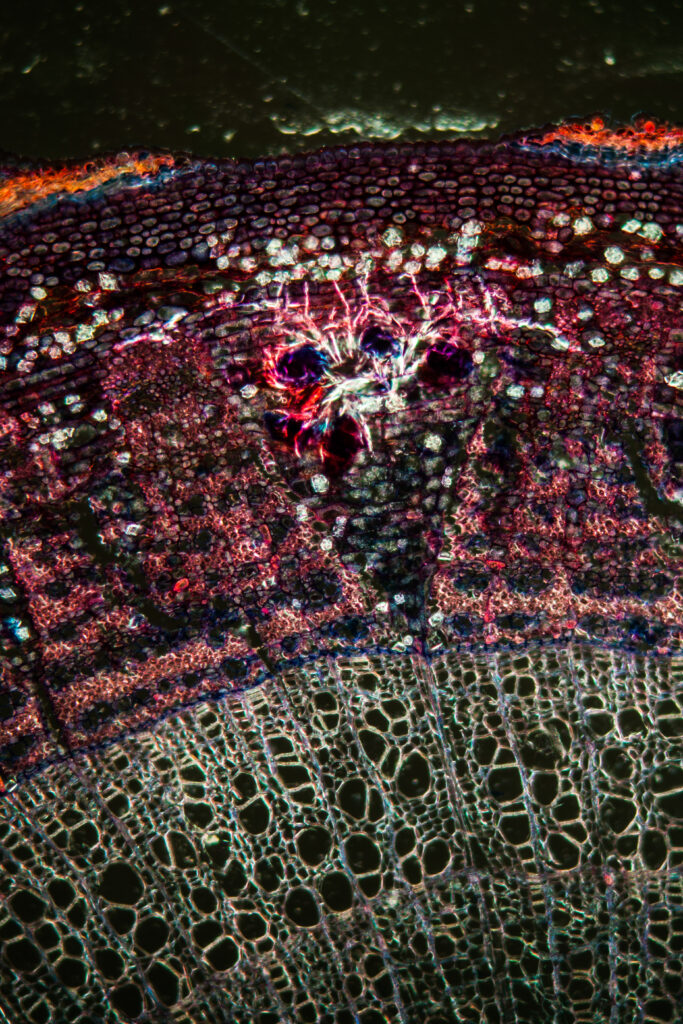
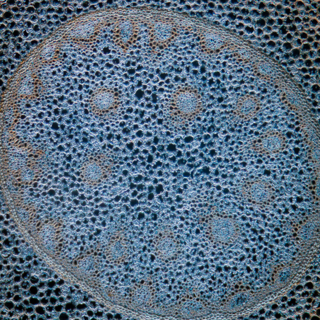
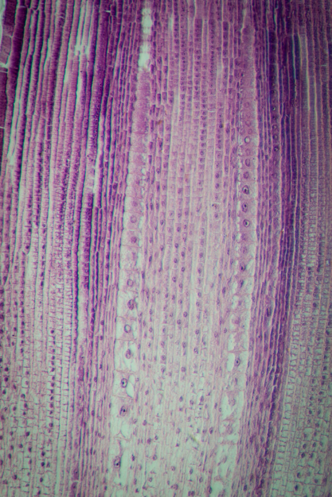
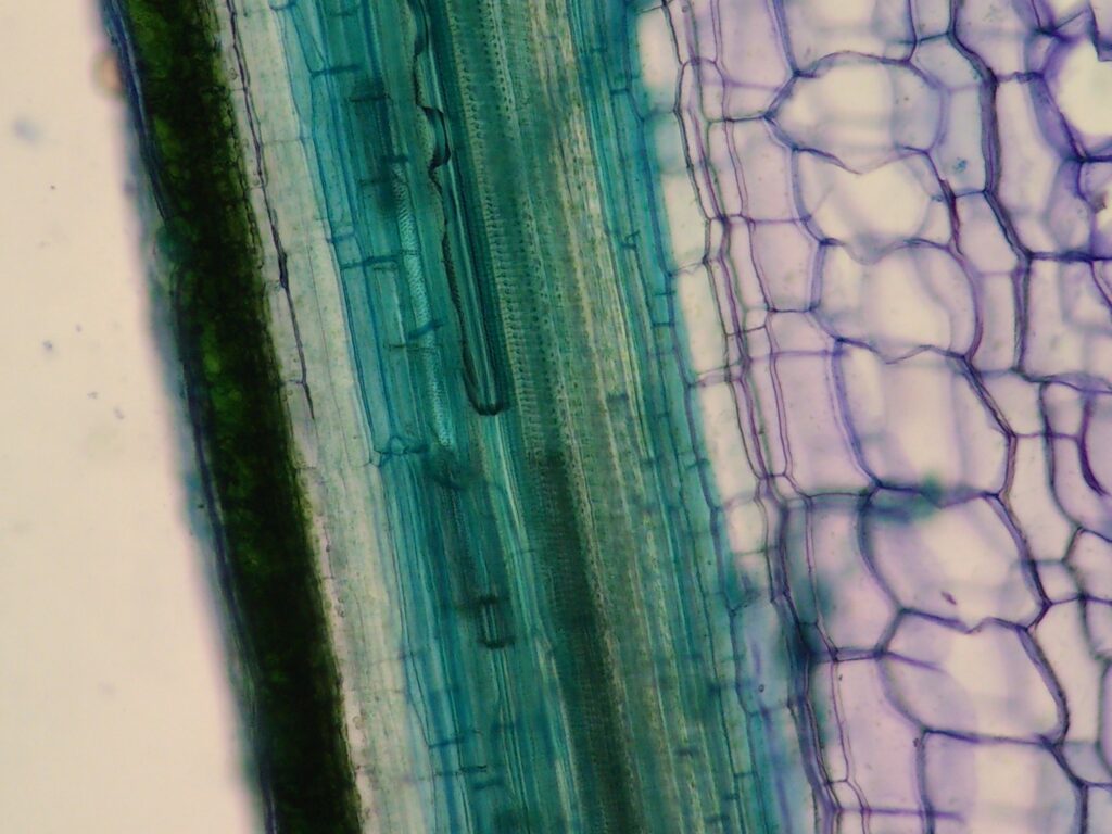
Oak
A cross section of a center (pith) portion of an older stem of Quercus (Oak). Magnification 100xA cross section of a Quercus (Oak) older stem showing typical structures. Magnification 40xPedunculate oak branch under the microscopePedunculate oak branch under the microscopeQuercus, oak, older woody root – microscopic cross section cut of a plant stem
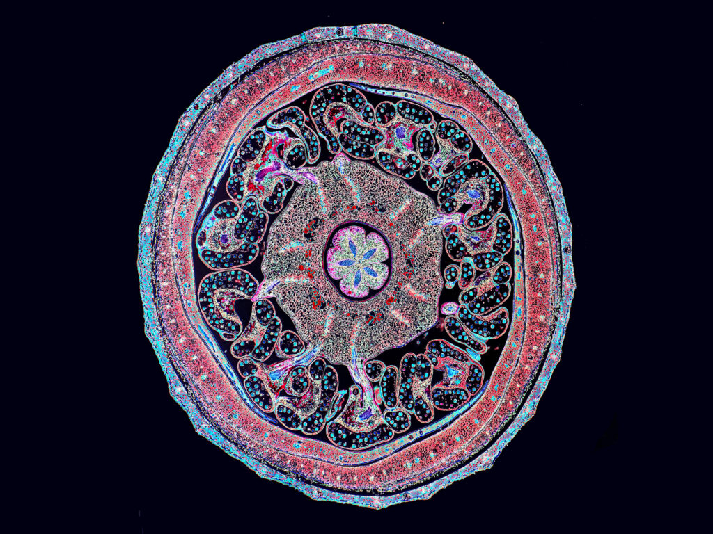
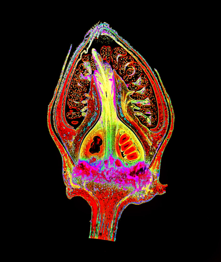
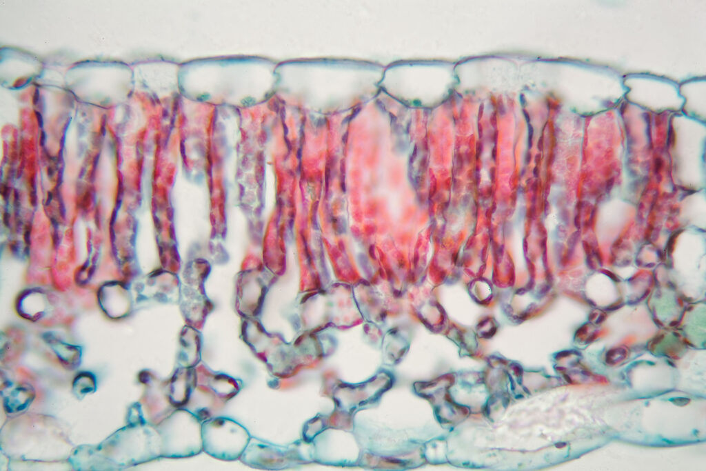
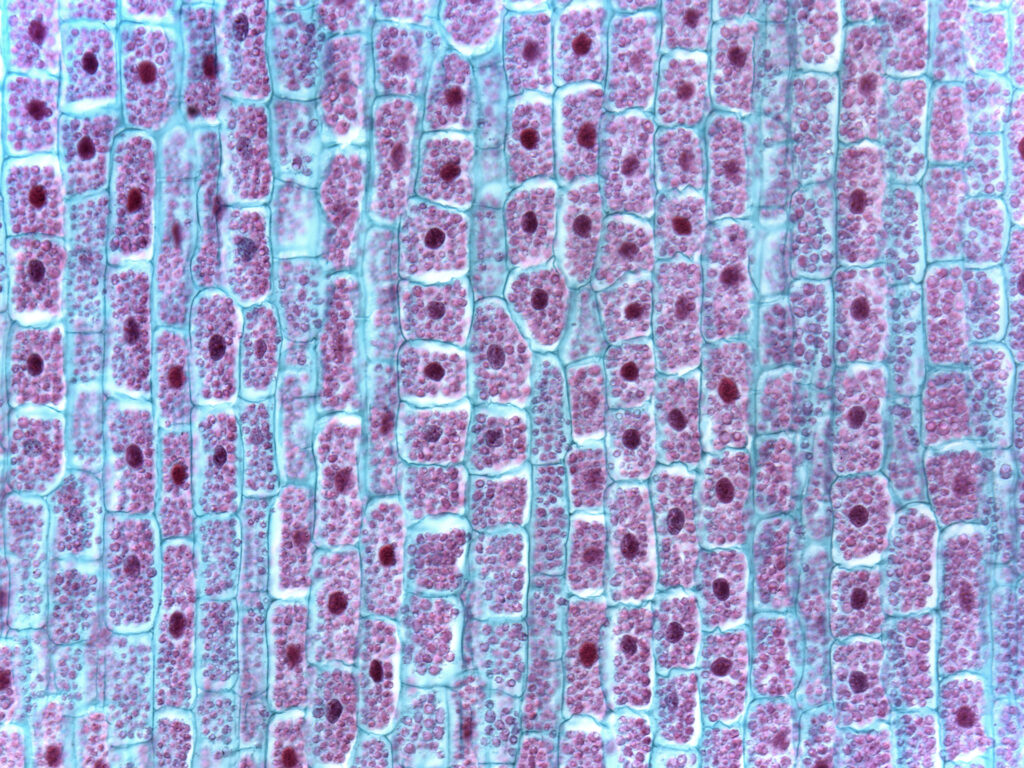
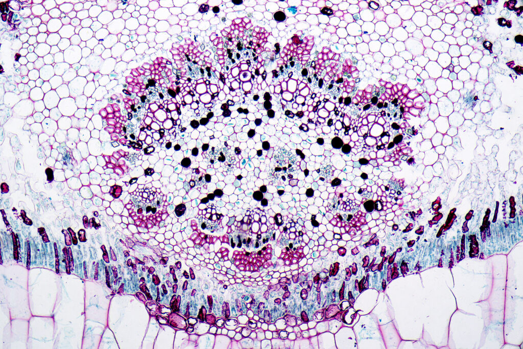
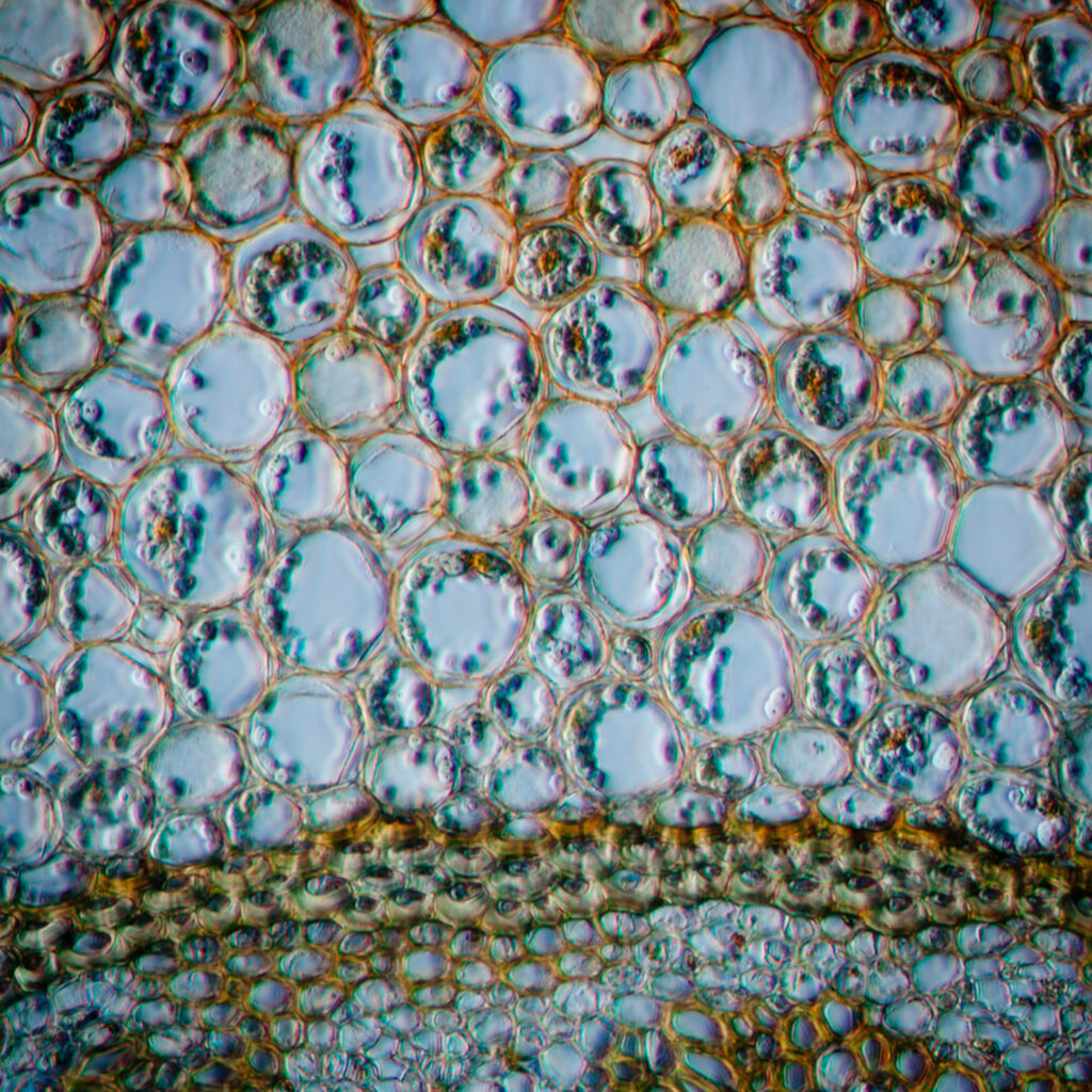
Cotton
A cross section of a floral bud of cotton (Gossypium) Image enhanced to show structural detail. Magnification 10xA cotton (Gossypium) fruit bud, longitudinal section, showing structure. Magnification 6x. Cross section of a cotton leaf under the microscope 2Cross section of a cotton leaf under the microscope
Maple
A cross section of the stem of a maple (Acer) tree. Magnification 40x. Image enhanced for structural detail.
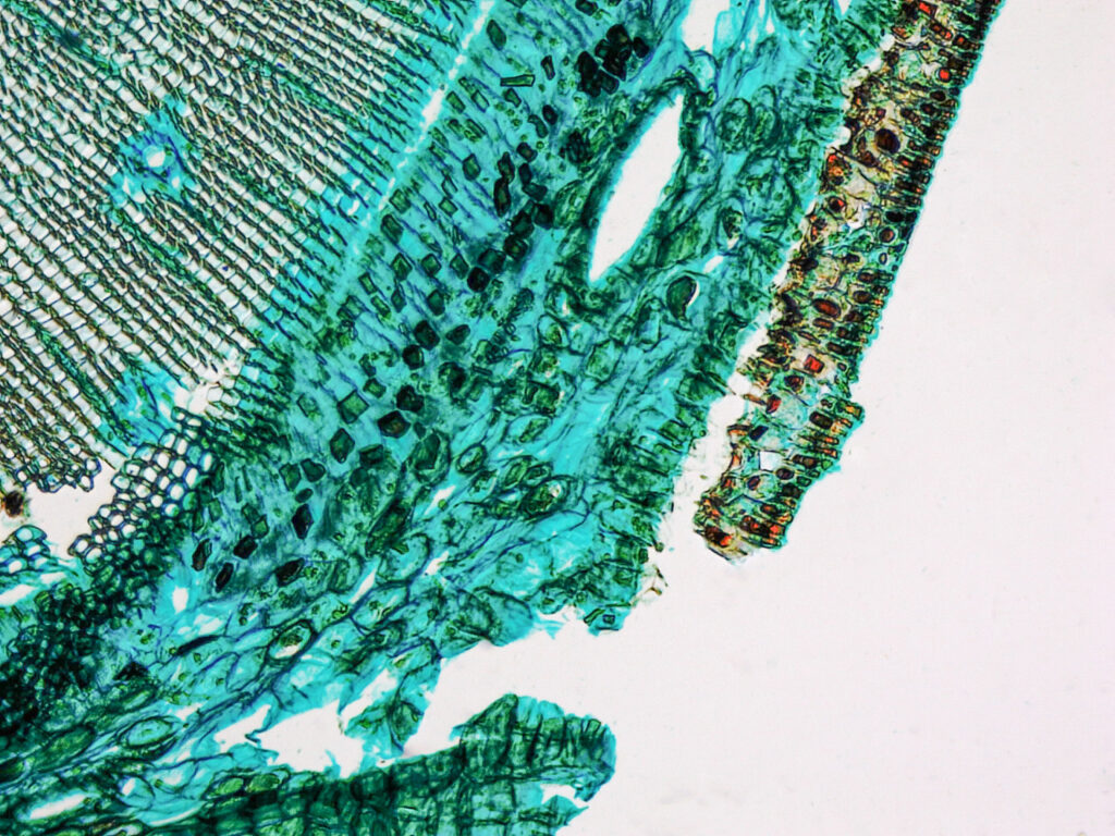
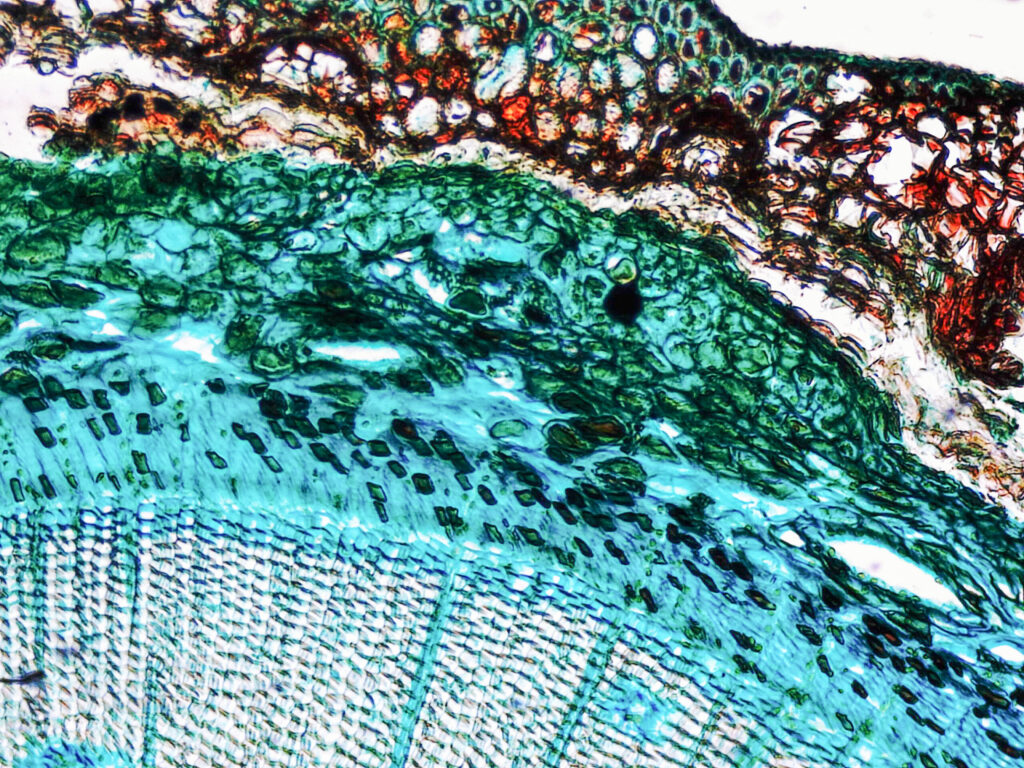
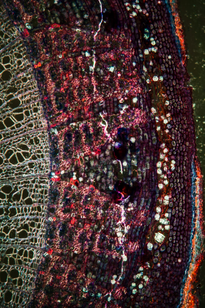
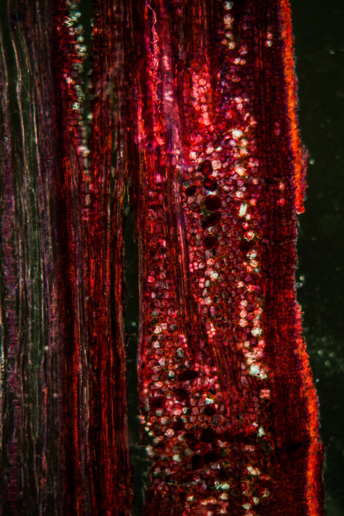
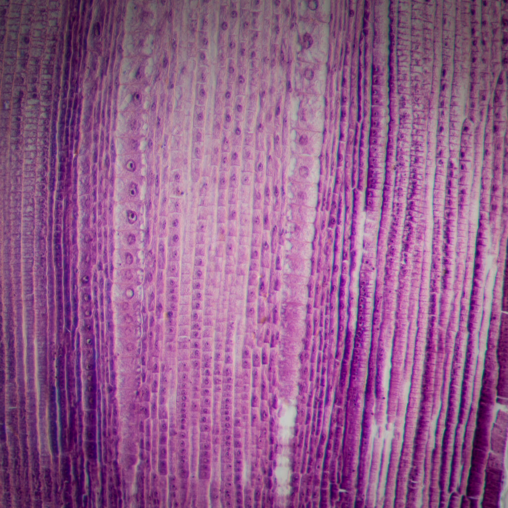
Cinnamon Bark
A detailed longitudinal section of cinnamon bark (Cinnamomum sp) showing associated structures and layers. Magnification 10x
Spruce (Needle)
Spruce needle under the microscope
Yew
Yew branch under the microscope
Larch
Larch branch under the microscopeLarch branch under the microscope
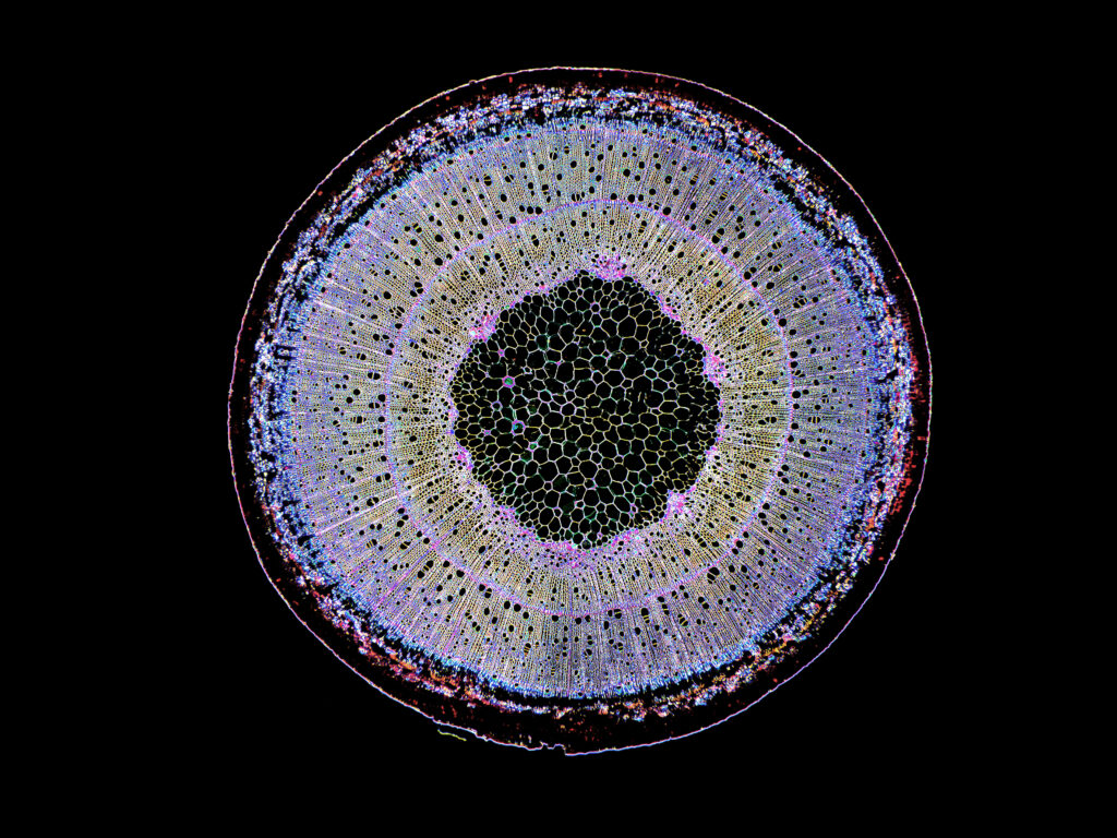
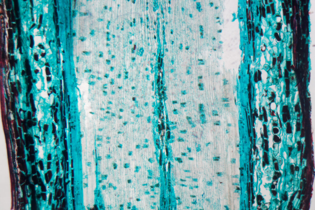
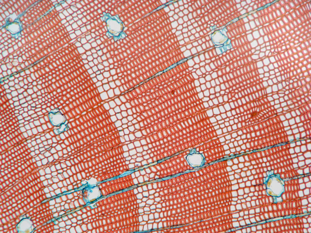
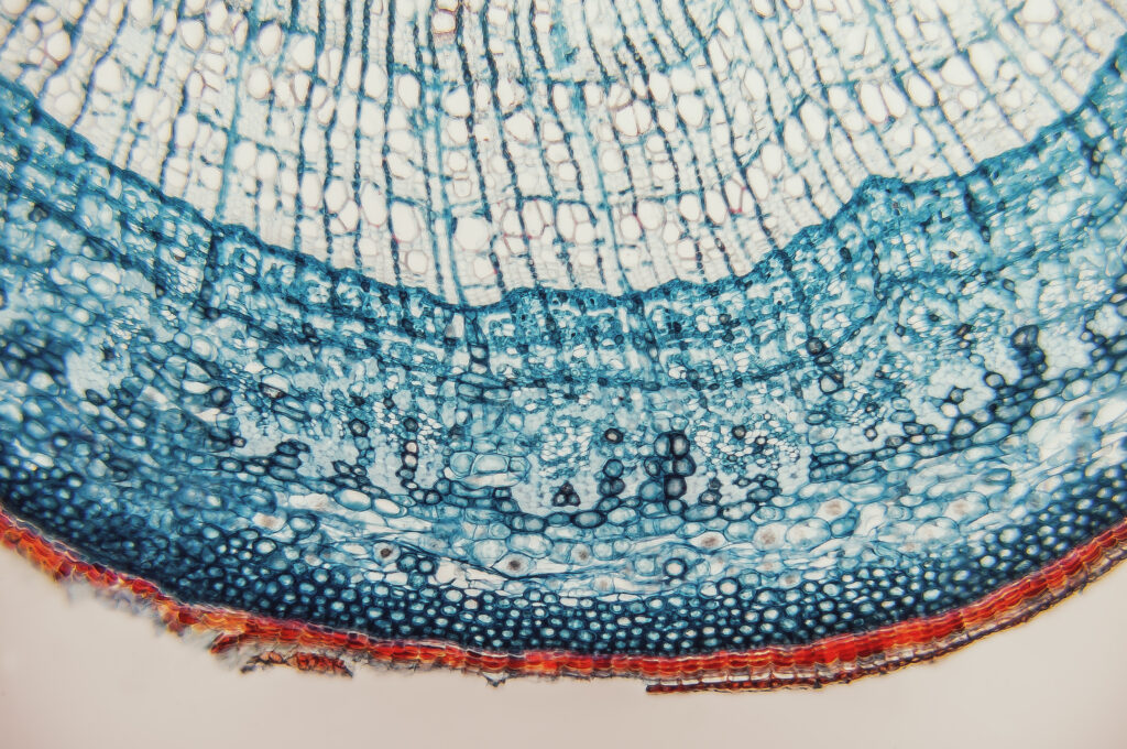
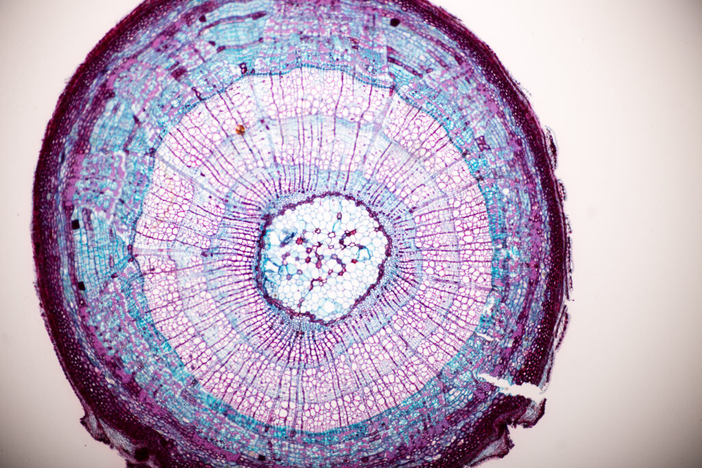
Pine
High resolution light photomicrograph of pine tree wood cross section seen through a microscopeHigh resolution light photomicrograph of pine tree wood cross section seen through a microscopeTransverse section of pine wood under microscope A section of pine wood under the microscope An scientific view of a female pine cone, longitudinal section, stained and mounted for microscopic examinationCrossection of pine needleLongitudinal section of a pine embryoResin ducts in a cross section of a pine stem. Magnification 100XCrossection of pine needleRoot of pine (Pinus) cross section. Magnification 40x. Image enhanced for detailPine Stem on the slide under microscope Pine treePine tree Pine Stem on the slide under microscope Transverse section of pine wood under microscope
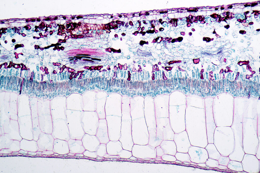
Rubber Plant
Rubber plant (Ficus elastica) leaf section under the microscopeRubber plant (Ficus elastica) leaf section under the microscope
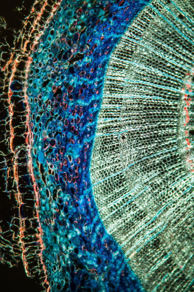
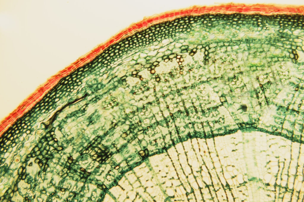
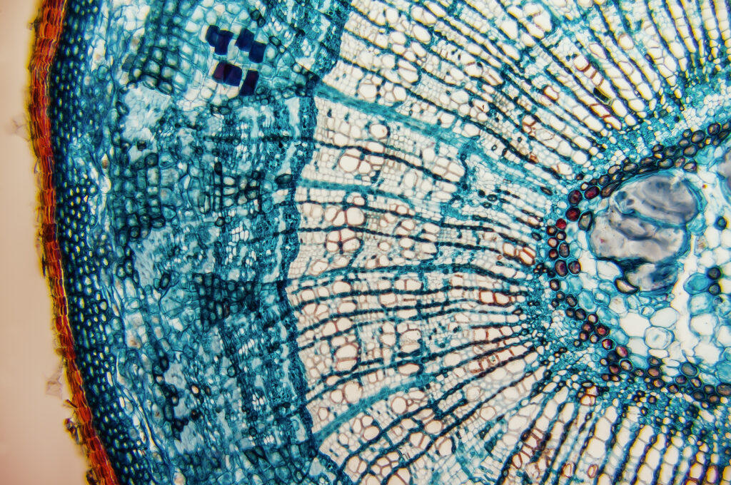
Basswood
High resolution light photomicrograph of tilia stem cross section seen through a microscopeHigh resolution light photomicrograph of tilia stem cross section seen through a microscopeTilia (Linden, Basswood) stem cross sectionCross section-cellular components wood cambium cells-heartwood and sapwood. Scientific research; plant tissue molecules magnification Duramen-formation of growth ringsCross section-cellular components wood cambium cells-heartwood and sapwood. Scientific research; plant tissue molecules magnification. Duramen- formation of growth ringsLight photomicrograph of tilia stem cross section seen through a microscopeThe internal structure cross section of tree- branches lindenLinden branch under the microscopeLinden branch under the microscope Linden branch under the microscopeThe internal structure cross section of tree- branches lindenLinden branch under the microscope
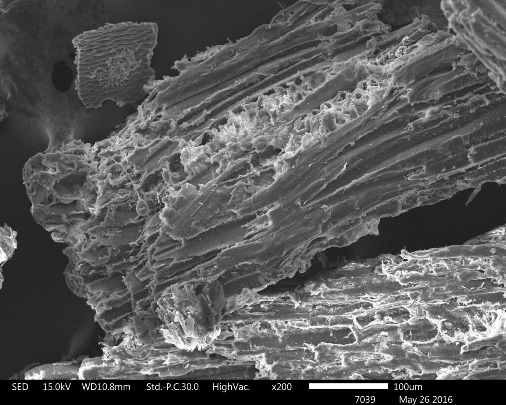
Wood Dust
Original scanning electron microscope images of wood sawdustscience botany micrograph plant root tissue background, 50 times magnifiedscience botany micrograph plant root tissue background, 100 times magnifiedscience botany micrograph plant root tissue background, 50 times magnifiedscience botany micrograph plant root tip tissue cell, Magnification 100Xscience botany micrograph plant root tip tissue cell, Magnification 100XPlant stem, long section under microscope viewcross-section branches or cut down a tree clearly distinguishes four layers the bark, cambium, wood coreCross section-Cellular components wood cambium cells-Heartwood and sapwood. Scientific research; plant tissue molecules magnification. Duramen-formation of growth ringsCross section-cellular components wood cambium cells- heartwood and sapwood. Scientific research; plant tissue molecules magnification.Duramen- formation of growth ringscambial cells- micro preparation samples- section (tissue) for microscope examinationCross-section Stem to show annual ring under the microscope
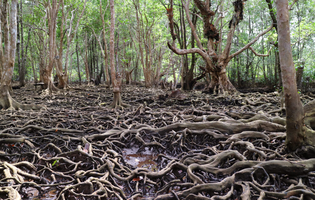
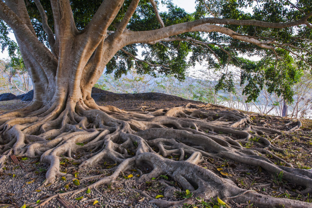
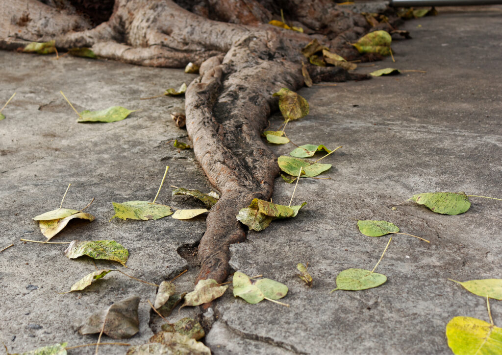
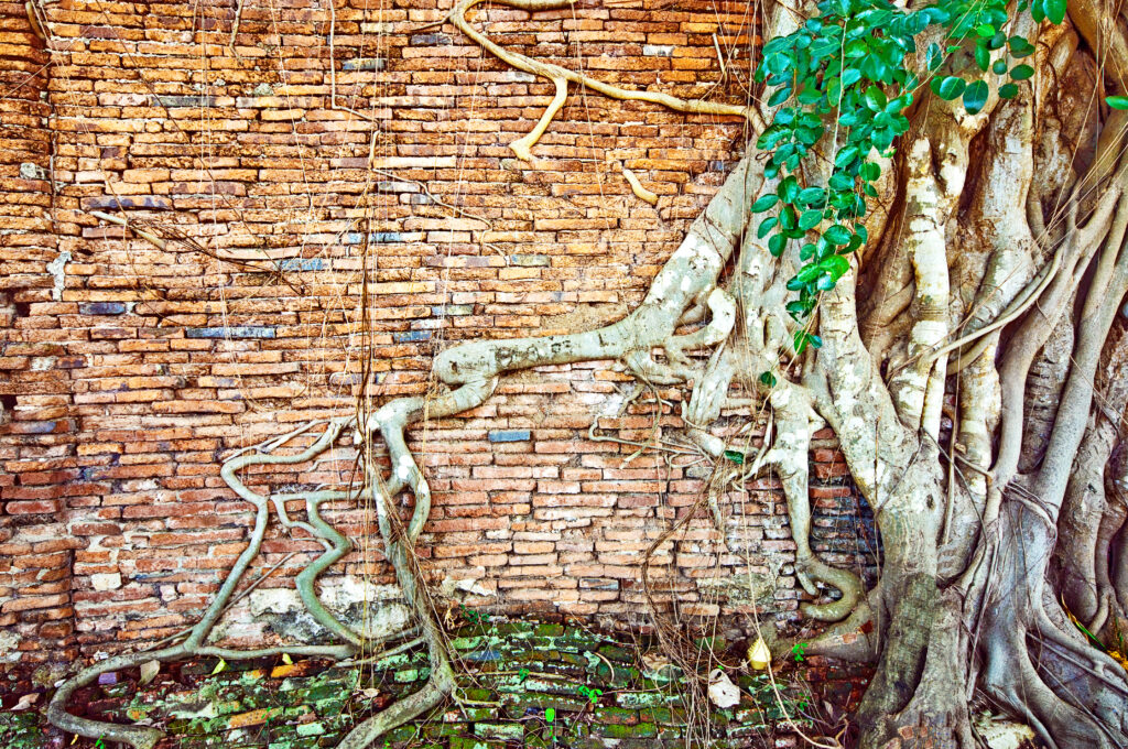
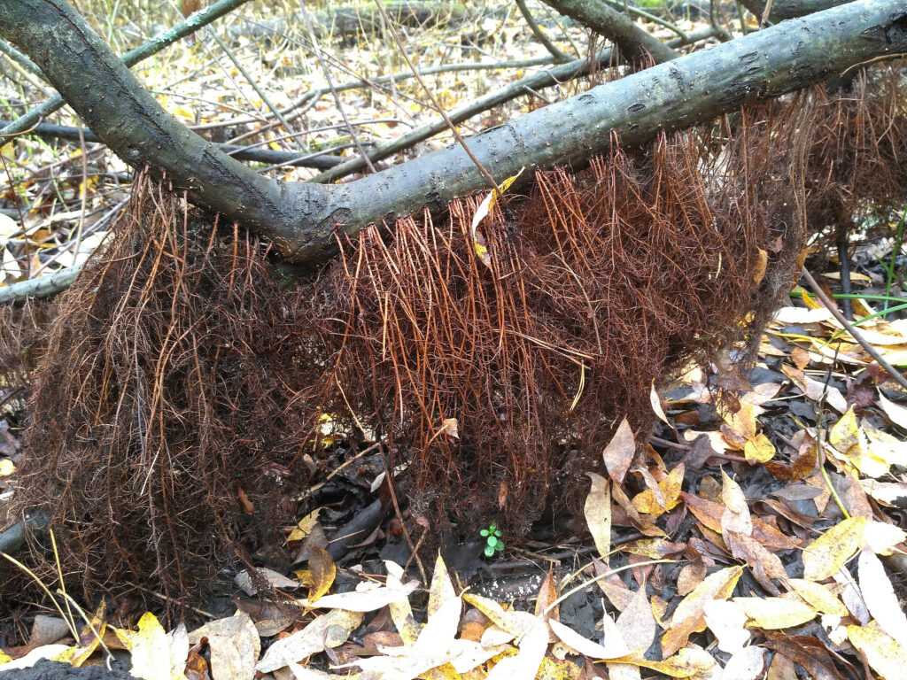
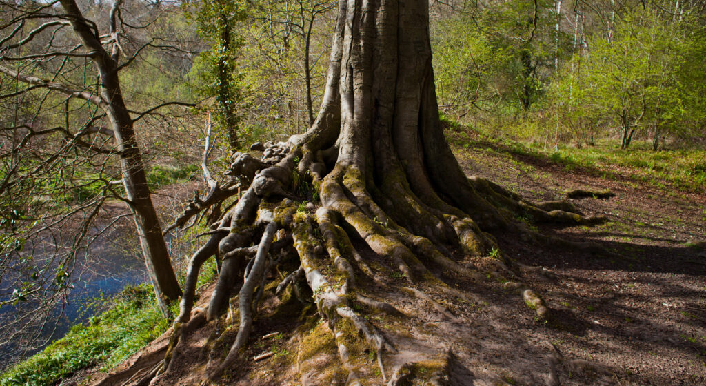
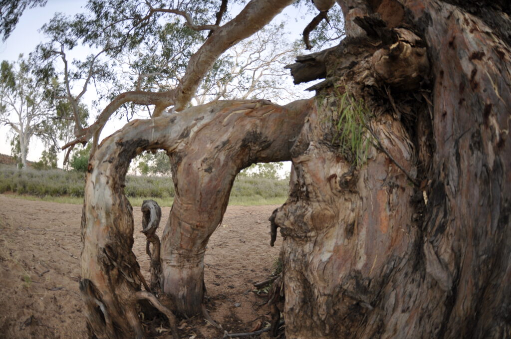
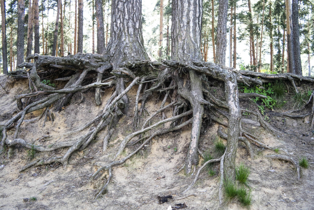
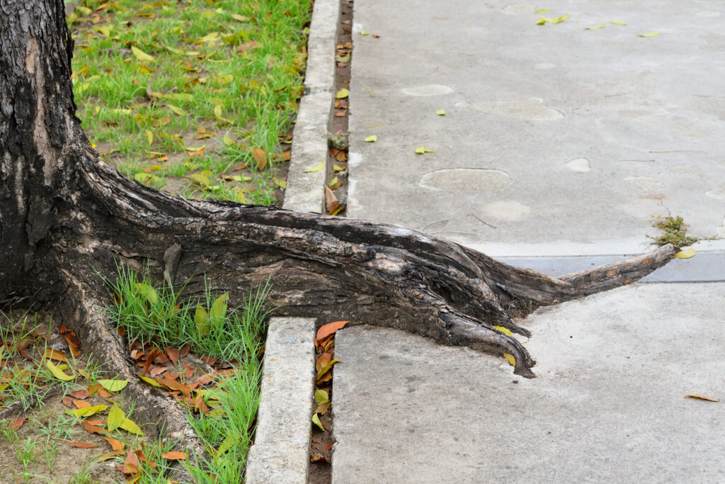
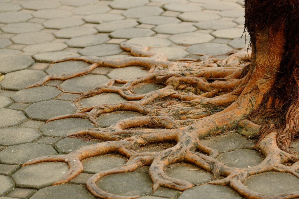
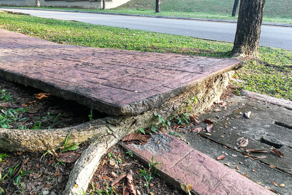
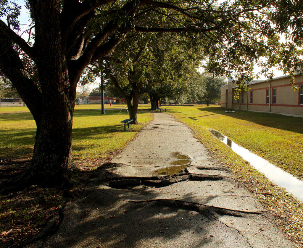
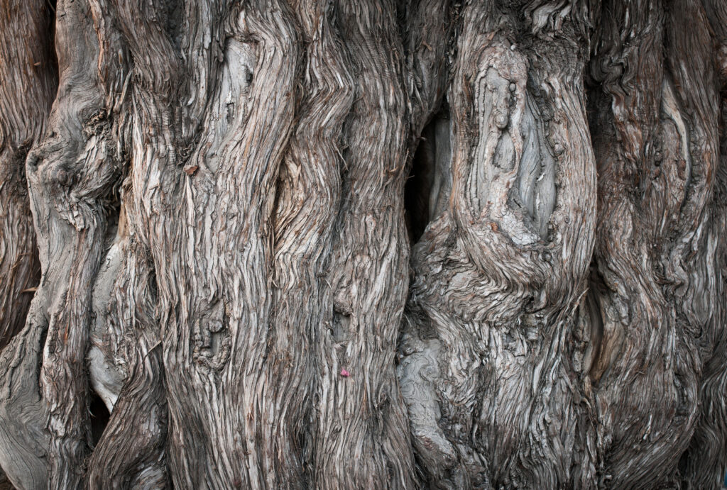
Root System
The Miracle of Gate way to the passage of time with arch is surrounded by bodhi Tree in Wat Phra Ngam ( Old buddhist temple ) Ayutthaya ,Thailand.Ayutthaya Buddha Head statue with trapped in Bodhi Tree roots at Wat Maha That (Ayutthaya). Ayutthaya historical park ThailandBig root of banyan tree land scape of ancient and old pagoda in history temple of Ayuthaya world heritage sites of unesco central of thailand important destinationBraided roots of large banyan tree in Sukhothai Historical Park, ThailandAmazing Tree Roots in the Mangrove Forest of Trat Province, ThailandRoot of the tree absorbing the ruins of the TempleRoots of a giant tree overgrowing the Temple Ta Prohm, Angkor, Cambodia, Asiaconcrete cover root of Sacred fig tree, leaf on floorOld brick wall with tree rootAccessory roots of the American maple (Acer negundo), denuded after lowering the water level of the forest lakeOld oak, tree rootsRiver Red Gum, Eucalyptus camaldulensis, East MacDonnell Ranges, Northern TerritoryTree roots inside of ancient wall in Ayutthaya Thailand